Aging, Cognition, & Memory Issues
Interested in learning how to keep your brain healthy and sharp as you age? Drs. Robert and Alanna Conder and I (Dr. Friesen) published a review article in OBM Geriatrics where we outline ways to promote healthy neuro-cognitive aging.
In our article we cover:
Click HERE for the link and HERE for the PDF.
At Niagara Neuropsychology, we offer 3 levels of assessment for Memory/Cognition/Early Dementia Assessment for Older Adults. These were developed in order to be able to provide the most comprehensive assessment possible and to ensure that even those with less financial means can obtain an adequate assessment.
Below are descriptions of each level of Memory/Cognition/Early Dementia Assessment for Older Adults from the top tier ADVANCED assessment, middle tier INTERMEDIATE assessment, and lowest tier BRIEF SCREENING assessment. We also offer repeat testing to determine or track change over time.
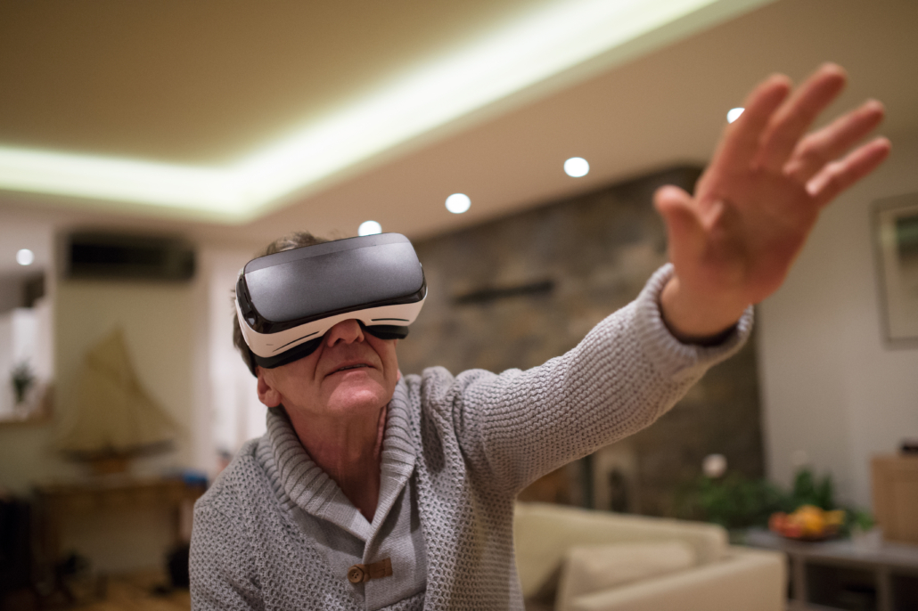
Virtual Reality Testing
Memory/Cognition/Early Dementia Assessment for Older Adults ADVANCED Assessment:
- The most advanced/thorough assessment of memory/cognitive and brain function
- A 55-minute intake interview with Dr. Friesen (charged separately at $250)
- Whole-brain quantitative electroencephalograph (qEEG)—brain mapping to measure brain function relative to others the same age and predict response to interventions like neurofeedback (~1-hour test)
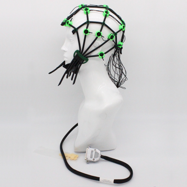
qEEG Brain Mapping
- Full neuropsychological and intellectual testing (up to age 97) to determine the presence and areas of cognitive impairment, including measuring learning, memory, visuospatial abilities, auditory processing, language, cognitive processing speed, attention, working memory, and executive functioning (~3-4 hours testing including Virtual Reality testing below)
- Virtual Reality Memory Testing (i.e., 360 degree and 3-D) up to age 90, uses a furniture store setting and measures immediate and delayed verbal and visual memory and learning (~30-minute test). See this video: https://youtu.be/iBwi0Gh_yc0
- Optional Virtual Reality Executive Functioning Testing (i.e., 360 degree and 3-D) u to 80, uses an ice-cream store setting which measures planning, working memory, processing speed, and cognitive flexibility( ~30-minute test). See this video: https://youtu.be/CmaqI2Arwew
- Optional Virtual Reality Attention Testing (i.e., 360 degree and 3-D). Continuous Performance Testing (CPT) is the gold standard for measuring selective and sustained visual and auditory attention, motor activity (hyperactivity), reaction time, and impulsivity (~30-minutes testing)
- Up to age 90, Virtual Reality testing uses an aquarium setting. See this video: https://youtu.be/2nc0WLxC7AY
- Full psychophysiological stress testing to determine and quantify problems with anxiety/coping/stress regulation (~30-minute test)
- Psychological testing for psychological/emotional difficulties (~60-90-minutes)
- Many aspects of memory/cognition naturally decline with age. This assessment helps determine whether a patient has normal age-related changes, mild cognitive impairment, pseudodementia (i.e., cognitive impairment due to depression) or more significant impairment as the result of various forms of dementia.
- If early signs of dementia are present, this assessment helps family physicians, neurologists, and/or geriatricians determine what the probable cause is (e.g., vitamin B-12 deficiency, vascular dementia, stroke, Alzheimer’s disease, Fronto-Temporal Dementia, Parkinson’s disease, traumatic brain injuries, concussions, etc.)
- Can re-test to track changes over time
- Brief report of test results provided
- 60-minute feedback session with Dr. Friesen to go over findings and treatment options
- Cost: $2050 (not including $250 intake session)
Memory/Cognition/Early Dementia INTERMEDIATE Assessment for Older Adults:
- Thorough and comprehensive assessment of memory/cognitive function
- A 55-minute intake interview with Dr. Friesen (charged separately at $250)
- Whole-brain quantitative electroencephalograph (qEEG)—brain mapping to measure brain function relative to others the same age and predict response to interventions like neurofeedback (~1-hour test)
- Targeted Neuropsychological and intellectual testing to determine the presence and areas of cognitive impairment, including measuring learning, memory, visuospatial abilities, auditory processing, language, cognitive processing speed, attention, working memory, and executive functioning (~2-3 hours testing including Virtual Reality testing below)
- Virtual Reality Memory Testing (i.e., 360 degree and 3-D) up to age 90, uses a furniture store setting and measures immediate and delayed verbal and visual memory and learning (~30-minute test). See this video: https://youtu.be/iBwi0Gh_yc0
- Psychological testing for psychological/emotional difficulties (~60-90-minutes)
- Many aspects of memory/cognition naturally decline with age. This assessment helps determine whether a patient has normal age-related changes, mild cognitive impairment, pseudodementia (i.e., cognitive impairment due to depression), or more significant impairment as the result of various forms of dementia.
- If early signs of dementia are present, this assessment helps family physicians, neurologists, and/or geriatricians determine what the probable cause is (e.g., vitamin B-12 deficiency, vascular dementia, stroke, Alzheimer’s disease, Fronto-Temporal Dementia, Parkinson’s disease, traumatic brain injuries, concussions, etc.)
- Can re-test to track changes over time
- Brief report of test results provided
- 60-minute feedback session with Dr. Friesen to go over findings and treatment options
- Cost: $1750 (not including $250 intake session)
Memory/Cognition/Early Dementia BRIEF SCREENING Assessment for Older Adults:
- A screening assessment of memory/cognitive function
- A 60-minute intake interview with Dr. Friesen (charged separately at $195)
- Basic neuropsychological and intellectual testing that includes measuring learning, memory, visuospatial abilities, auditory processing, language, cognitive processing speed, attention, working memory, and executive functioning (~90-minutes testing)
- Psychological testing for psychological/emotional difficulties (~45-minutes)
- Determines presence and areas of cognitive impairment
- Many aspects of memory/cognition naturally decline with age. This assessment helps determine whether a patient has normal age-related changes, mild cognitive impairment, pseudodementia (i.e., cognitive impairment due to depression), or more significant impairment as the result of various forms of dementia
- If early signs of dementia are present, this assessment helps neurologists and geriatricians determine what the probable cause is (e.g., vitamin B-12 deficiency, vascular dementia, stroke, Alzheimer’s disease, Fronto-Temporal Dementia, Parkinson’s disease, traumatic brain injuries, concussions, etc.)
- Can re-test to track changes over time
- 60-minute feedback session with Dr. Friesen to go over findings and treatment options
- Brief summary report provided
- Cost: $1350(not including $250 intake session)
NORMAL AGING VS. DEMENTIA
*Adapted from the Alzheimer Society Canada
Alzheimer’s disease and other dementias are not a part of normal aging.
Almost 40 percent of people over the age of 65 experience some form of memory loss. When there is no underlying medical condition causing this memory loss, it is known as “age-associated memory impairment,” which is considered a part of the normal aging process.
Brain diseases like Alzheimer’s disease and other dementias are different.
Age-associated memory impairment and dementia can be told apart in a number of ways. Below are some examples.
Note: this is not a diagnostic tool.
| Normal Aging | Dementia |
| Not being able to remember details of a conversation or event that took place a year ago | Not being able to recall details of recent events or conversations |
| Not being able to remember the name of an acquaintance | Not recognizing or knowing the names of family members |
| Forgetting things and events occasionally | Forgetting things or events more frequently |
| Occasionally have difficulty finding words | Frequent pauses and substitutions when finding words |
| You are worried about your memory but your relatives are not | Your relatives are worried about your memory, but you are not aware of any problems |
If you are worried about your memory, talk to your family doctor.
Tips for coping with normal age-related memory difficulties:
- Keep a routine
- Organize information (keep details in a calendar or day planner)
- Put items in the same spot (always put your keys in the same place by the door)
- Repeat information (repeat names when you meet people)
- Run through the alphabet in your head to help you remember a word
- Make associations (relate new information to things you already know)
- Involve your senses (if you are a visual learner, visualize an item)
- Teach others or tell them stories
- Get a full night’s sleep
- Learn more about what you can do to maintain your brain health and strengthen your memory
WHAT IS DEMENTIA?
Dementia is an overall term for a set of symptoms that are caused by disorders affecting the brain. Symptoms may include memory loss and difficulties with thinking, problem-solving or language, severe enough to reduce a person’s ability to perform everyday activities. A person with dementia may also experience changes in mood or behaviour.
Dementia is generally considered to be progressive, which means the symptoms will gradually get worse as more brain cells become damaged and eventually die.
Dementia is not a specific disease. Many diseases can cause dementia, including Alzheimer’s disease, vascular dementia (due to strokes), Lewy Body disease, head trauma, fronto-temporal dementia, Creutzfeldt-Jakob disease, Parkinson’s disease, and Huntington’s disease. These conditions can have similar and overlapping symptoms.
Some treatable conditions can produce symptoms similar to dementia, for example, vitamin deficiencies, thyroid disease, sleep disorders, or psychiatric/psychological disorders. It is therefore important to arrange for a full medical assessment as early as possible.
Getting a timely diagnosis can help you access information, resources, and support through the Alzheimer Society, benefit from treatment, and plan ahead.
GETTING A DIAGNOSIS
Finding out if it’s dementia
Alzheimer’s disease and other dementias are not a normal part of aging. This list of 10 warning signs will help you understand if you should seek further information.
Other conditions have symptoms similar to dementia and may be treatable, including depression, chest and urinary infections, severe constipation, vitamin and thyroid deficiencies and brain tumours, drug interactions or alcohol abuse. Other possible causes of confusion are poor sight or hearing; and emotional changes and upsets, such as moving or bereavement. Finding out the cause of the symptoms can help the person get the proper care, treatment and support.
If you are concerned about any symptoms, go to your doctor. If you don’t receive the help you need, ask to be referred to a specialist. You know your body and you should speak up if you have worries.
To learn more about what to expect during the Alzheimer diagnosis process, review our brochure on getting a diagnosis.
The diagnostic process: assessments and tests
The diagnostic process begins with your family doctor. After your initial assessment, he/she should refer you to a specialist like a neurologist, psychiatrist, geriatrician, or neuropsychologist. Your family physician may recommend additional testing. After a diagnosis is made, you will likely continue to see your family doctor for ongoing assessment.
Initial assessment
During your initial assessment, your family doctor will ask you questions about your symptoms, past illnesses, and family medical and psychiatric history. You may also undergo physical examinations and tests, which may include detailed blood work to look for heart, lung, liver, kidney or thyroid problems that may be causing your symptoms. To evaluate whether another nervous system disorder may be causing the symptoms, your doctor may also test muscle tone and strength, coordination, eye movement, speech and sensation.
Your doctor may also conduct mental tests to measure your sense of time and place as well as your ability to remember, express yourself, and perform simple calculations. This may involve exercises such as recalling words and objects, drawing and spelling, and questions such as “What year is it?” After an initial assessment, your doctor may feel able to make a diagnosis, but should generally refer you to a memory clinic or other specialist service for more testing.
Additional tests
Other tests such as X-rays and EEGs (electroencephalograms) may be used to determine the source of the problem. In some centres, scans may be used. The following may be recommended, but are not always necessary for a diagnosis:
- CT (computerized tomography) scan and MRI (magnetic resonance imaging) to take images of the brain.
- SPECT (single proton emission computed tomography) showing how blood is circulating to the brain.
- PET (positive electron tomography) showing how the different areas of the brain respond during certain activities, for example, reading and talking.
Referral to a specialist
If you feel that a referral would be helpful and your family doctor does not suggest it, you can request it. Your doctor may refer you to one of the following specialists:
- Neurologist, who specializes in disorders of the brain and nerve pathways. Some neurologists have particular experience in diagnosing dementia.
- Geriatrician, who specializes in the physical illnesses and disabilities associated with old age and in the care of older people.
- Psychiatrist, who specializes in diagnosing and treating a wide range of mental health problems. A psychiatric evaluation may be helpful in ruling out other illnesses such as depression, which can cause symptoms similar to those associated with Alzheimer’s disease. Geriatric psychiatrists are psychiatrists who have further specialized in the mental health problems of older people, including dementia.
- Clinical Neuropsychologist (Dr. Friesen is a clinical neuropsychologist), who specializes in assessing and treating normal and abnormal functioning of the central nervous system (i.e., brain). Clinical neuropsychologists address neurobehavioral problems related to acquired or developmental disorders of the nervous system. The types of problems are extremely varied and include such conditions as dementia, vascular disorders, Parkinson’s disease and other neurodegenerative disorders, traumatic brain injury, seizure disorders, learning disabilities, neuropsychiatric disorders, infectious disease affecting the CNS, neurodevelopmental disorders, metabolic disease and neurological effects of medical disorders or treatment. Clinical neuropsychologists apply specialized knowledge in the assessment, diagnosis, treatment and rehabilitation of individuals with neurological, medical, or neurodevelopmental disorders across the lifespan.
Ongoing assessment
Once a diagnosis is confirmed, make an appointment to see your family doctor from time to time to assess changes and discuss any problems. Your doctor may refer you to a specialist, such as a neuropsychologist for help in assessing changes, and for advice on ways to deal with specific difficulties. Your family doctor is also responsible for your general health when you have dementia.
ALZHEIMER’S DISEASE
At this point in time, Alzheimer’s disease is irreversible and destroys brain cells, causing thinking ability and memory to deteriorate. Alzheimer’s disease is not a normal part of aging.
What is Alzheimer’s disease?
Dr. Alois Alzheimer first identified the disease in 1906. He described the two hallmarks of the disease: “plaques,” which are numerous tiny, dense deposits scattered throughout the brain that become toxic to brain cells at excessive levels, and “tangles,” which interfere with vital processes, eventually choking off the living cells. When brain cells degenerate and die, the brain markedly shrinks in some regions.
The image below shows that a person with Alzheimer’s disease has less brain tissue (right) than a person who does not have the disease (left). This shrinkage will continue over time, affecting how the brain functions.

MRI images courtesy of Sunnybrook and
Women’s College Health Sciences Centre
What is the difference between Alzheimer’s disease and dementia?
If you have been confused by these terms in the past, or mistakenly thought that they were the same thing, watch the video:
The material was created by TCD, through the NEIL Programme at the Institute of Neuroscience with support from GENIO.
The effects of Alzheimer’s disease
Alzheimer’s disease is a fatal disease that eventually affects all aspects of a person’s life: how they think, feel, and act. Each person is affected differently. It is difficult to predict symptoms, the order in which they will appear, or the speed of their progression.
The following are some of the changes you may expect as the disease progresses.
Cognitive and functional abilities: a person’s ability to understand, think, remember and communicate will be affected. This could impact a person’s ability to make decisions, perform simple tasks, or follow a conversation. Sometimes people lose their way, or experience confusion and memory loss, initially for recent events and eventually for long-term events.
Emotions and moods: a person may appear apathetic and lose interest in favourite hobbies. Some people become less expressive and withdrawn.
Behaviour: a person may have reactions that seem out of character. Some common reactions include repeating the same action or words, hiding possessions, physical outbursts and restlessness.
Physical abilities: the disease can affect a person’s coordination and mobility, to the point of affecting their ability to perform day-to-day tasks such as eating, bathing and getting dressed.
Resources:
Risk factors – how can we reduce the risk?
10 warning signs – know the signs and symptoms
What is Alzheimer’s disease? – brochure by the Alzheimer Society of Canada
Stages of Alzheimer’s disease – learn about the progression of the disease
Shared experiences – advice from people living with Alzheimer’s disease
OTHER DEMENTIAS
“Other dementias” resemble Alzheimer’s disease in that they also involve a progressive degeneration of brain cells that is currently irreversible. There are many different types of dementia, although some are far more common than others.
- Young onset dementia
- Mild Cognitive Impairment
- Vascular dementia
- Mixed dementia
- Frontotemporal dementia
- Lewy body dementia
- Creutzfeldt-Jakob disease
- Down syndrome
- Rarer forms of dementia
A small percentage of dementias are reversible, occurring as a secondary development in treatable conditions. Toxic reactions to prescription or over the counter medications are the most common cause of reversible dementia. Others include dietary or vitamin B12 deficiencies, infections, tumours, alcoholism, inflammatory states, hormonal dysfunction, environmental toxins, drug abuse, and depression.
TREATMENT OPTIONS
There is currently no cure for Alzheimer’s disease and other dementias, nor is there any known treatment that will stop the progression. Several drugs on the market and non-pharmacological treatments may help with some symptoms.
Some non-pharmacological therapies (such as music therapy, neurofeedback, transcranial photobiomodulation, tDCS) may be beneficial to people with dementia. However, at this point in time, much of the research, although appearing promising, is still in its infancy which prevents us from determining its effectiveness. We at Niagara Neuropsychology now offer neurofeedback and photobiomodulation, to those experiencing cognitive impairment. See a recent review of the use of photobiomodulation in the treatment of Alzheimer’s disease HERE and HERE.
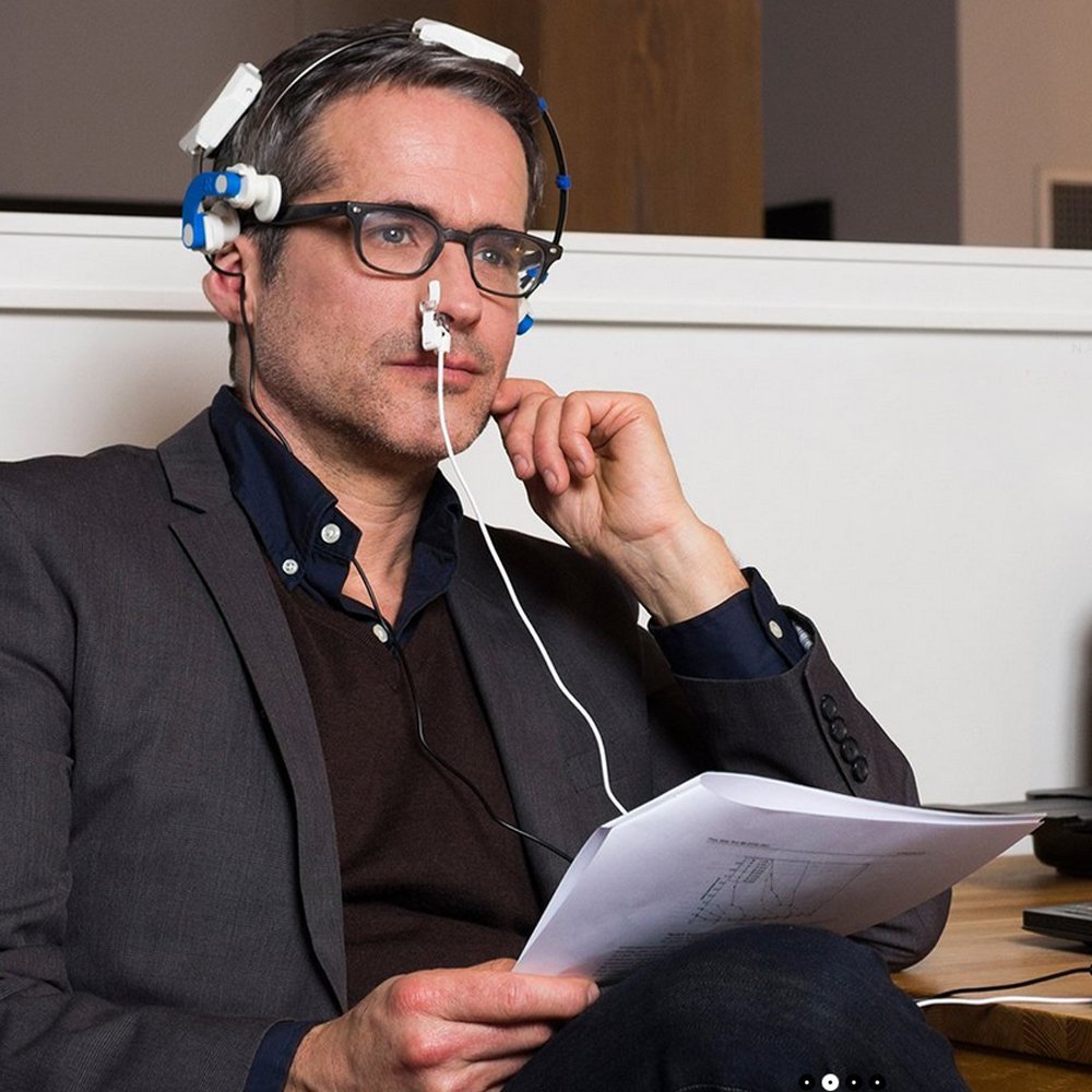
VieLight Neuro
Transcranial Photobiomodulation (aka Neurophotomodulation) or low-level light therapy (LLLT) involves the use of low-level near-infrared light to stimulate neuronal mitochondria and cellular events. These lights are applied to the patient’s head for therapeutic purposes such as recovery from neurological disorder or damage. Light in the red and near-infrared regions of the electromagnetic spectrum are used because of their ability to penetrate the scalp, skull, and brain. Studies have been performed to assess the safety and effectiveness of transcranial light therapy in conditions such as depression, stroke, traumatic brain injury (TBI), and neurodegenerative conditions.
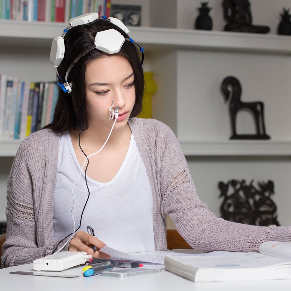
VieLight Neuro
For a review of the research, click here (general review), here , here, here , here, here, and here (Alzheimer’s and Parkinson’s disease), here , here (TBI), here(mild TBI/concussions) here, here (improved cognitive and mood in healthy subjects), here(reduction in depression in patients with Major Depressive Disorder) and here (references).
The first study to report significant cognitive improvement in dementia participants following brain photobiomodulation treatments was presented at the Alzheimer’s Association International Conference in Toronto in July 2016. To see the abstract, click here. To see the poster, click here. To see a related news clip click here.
Due to the fact that only recently have researchers started to use transcranial photobiomodulation in the treatment of brain disorders and, although transcranial photobiomodulation is considered safe, at this point in time it is considered experimental and thus no medical claims can be made.
The International Society for Neuroregulation & Research (ISNR.org) regularly updates a comprehensive bibliography of neurofeedback research studies on various disorders including cognitive decline and enhancement that can be accessed HERE.
When considering the use of natural health products, think about the following to minimize your risk:
- Don’t assume “natural” means “safe.”
- Be wary of unsubstantiated health-related claims.
- Herbal remedies can change the way prescription drugs work. Be aware of interactions with other drugs and tell your doctor and pharmacist about any herbal remedies you may be taking.
Additional resources:
Heads Up for Healthier Living: information on lifestyle choices that can improve the quality of life for people living with dementia and may help to slow the progression of the disease.
Information sheets on the following medications, including possible benefits and side effects:
- Reminyl®
- Rivastigmine (Exelon®)
- Aricept®
- Memantine hydrochloride (Ebixa®)
PARKINSON’S DISEASE
We will have more to add to this section in the future. For now, there is some interesting work being done with neurofeedback and Parkinson’s disease. To learn more, click here:
BRAIN HEALTH
The human brain is one of your most vital organs. It plays a role in every action and every thought, and just like the rest of your body, it needs to be looked after.
Can Alzheimer’s disease be prevented? There are no guarantees, but healthy lifestyle choices will help keep your brain as healthy as possible as you age.
By making better lifestyle choices now, you can improve your brain’s ability to sustain long-term health and fight illnesses.
Be good to your brain: